
Turn-Key Spatial Biology
Turn complex multiplex slides into standardized, cell-level answers - without a bioinformatics pipeline.
PhenoScout reads all major mIF file formats and brightfield images. mIF Assays are analysed with one pre-trained AI Foundation Model, identifying and quantifying phenotypes, and unlocking spatial context at scale, straight out of the box.

No Pipeline to Build. Pre-trained models, ready on upload

Reproducible at Scale. Consistent reads across sites, scanners and operators

Spatial Context, Decoded. Single-cell phenotypes and tissue neighborhoods
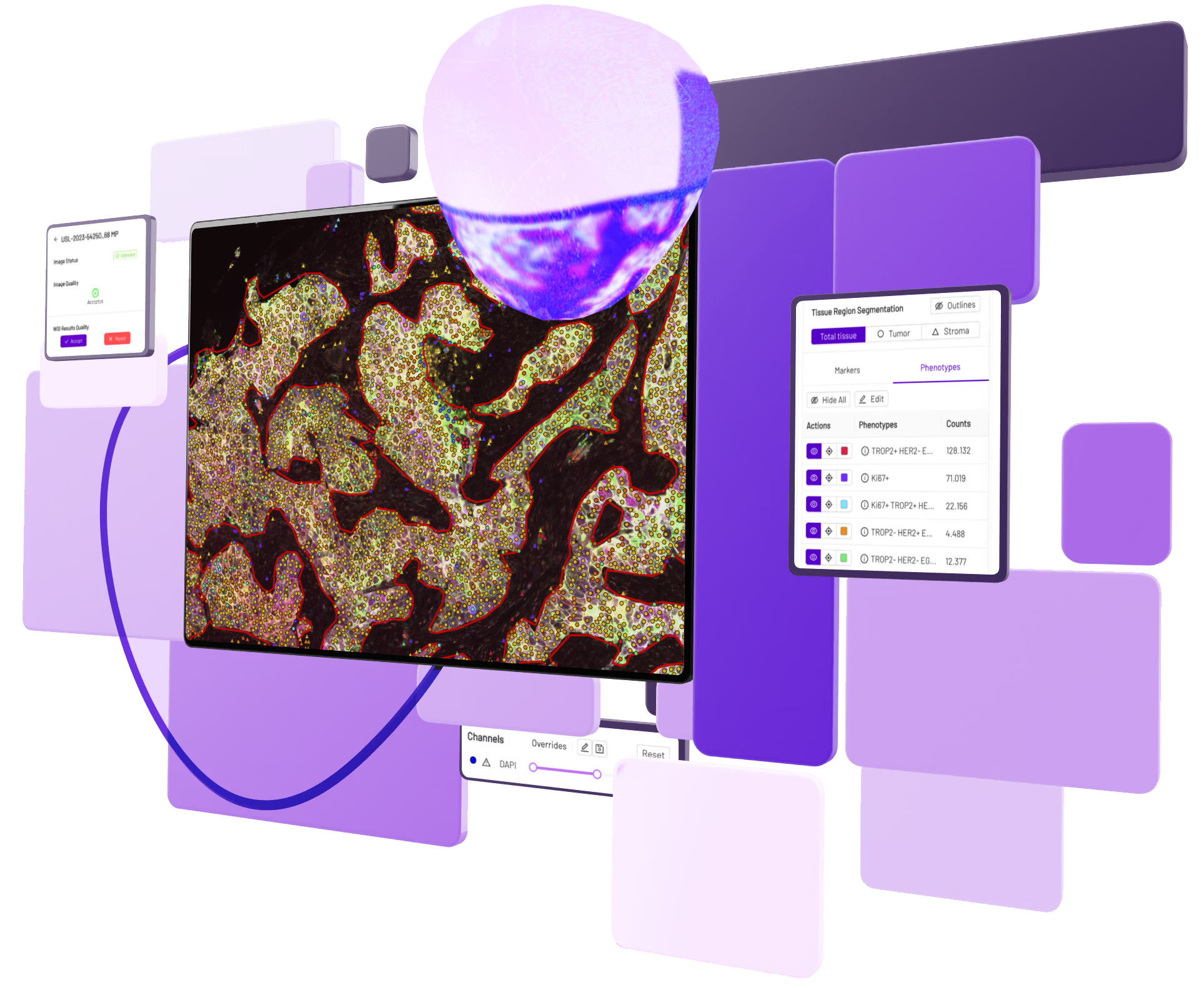
From Multiplex to Meaning.
PhenoScout turns routine multiplex immunofluorescence slides into standardized, quantitative spatial biology - analyzed in a fully automated cloud workflow.
Built on Mindpeak's patented peakFrontier foundation model, PhenoScout detects cells and classifies phenotypes in mIF images, removing the manual bottlenecks that have kept spatial biology out of routine use.
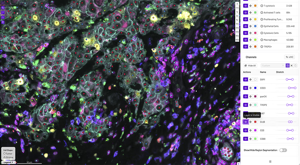
State-of-the-Art AI for Standardized Multiplex Tissue Analysis
Spatial biology has transformed discovery - but once imaging is complete, analysis becomes the bottleneck:
mIF images are beyond human readability for visual scoring.
Too many markers, too much signal, too little time.
Success depends on a tight assembly line across three scarce disciplines.
Pathologists for QC and annotation, image scientists for cell detection and segmentation, bioinformaticians for spatial readouts.
Every image adds QC workload, storage and processing burden
and analysis results are highly complex: marker combinations, spatial relationships, explainable outputs.
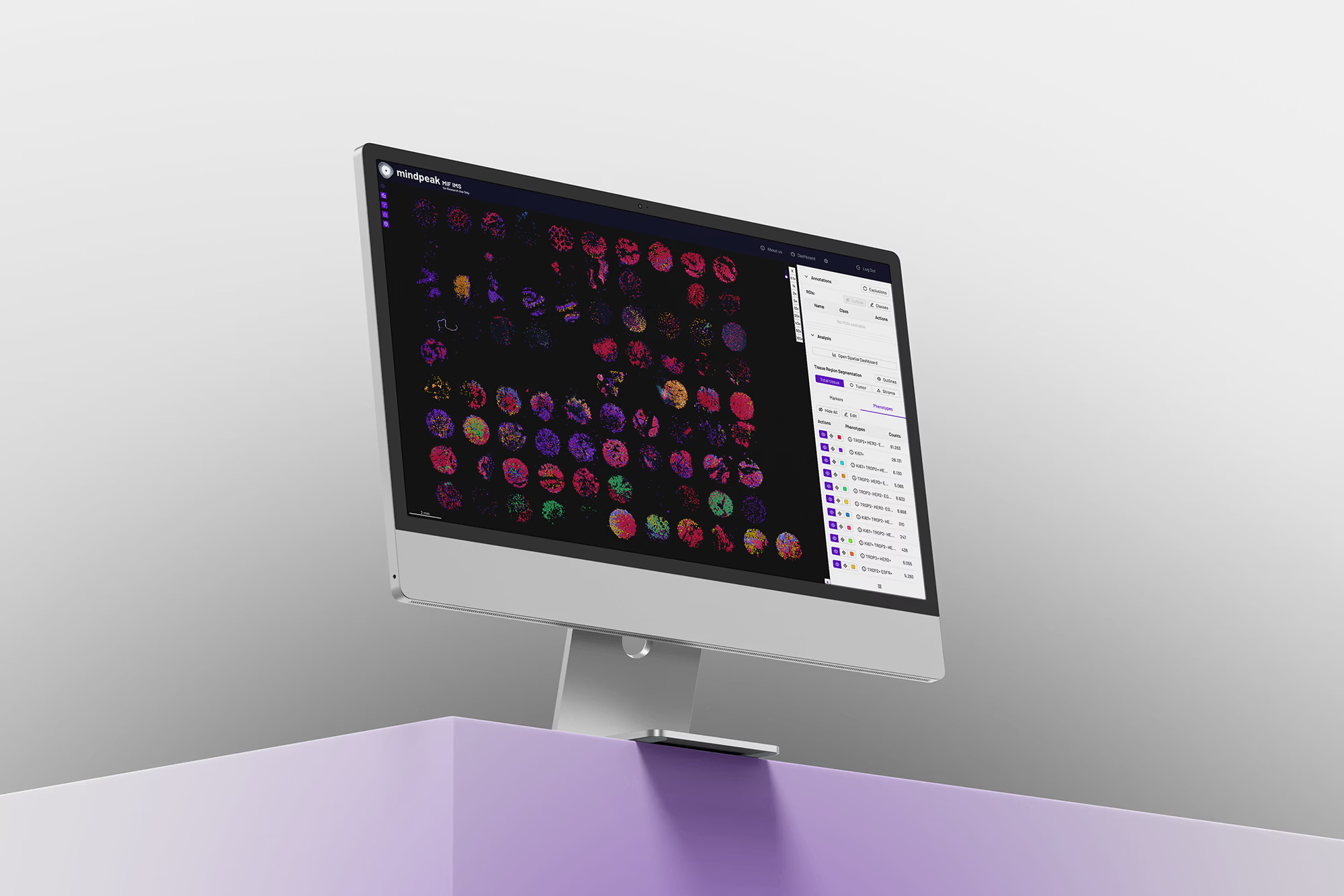
PhenoScout answers these directly from the multiplex and brightfield slides you already generate.
No custom pipeline.
No specialized image-analysis or bioinformatics expertise.
No waiting in the bioinformatics queue.
The platform integrates, spatial and cellular data to deliver fast, reproducible, standardized readouts
Supporting biomarker discovery, patient stratification and translational research across oncology and immunology pipelines.
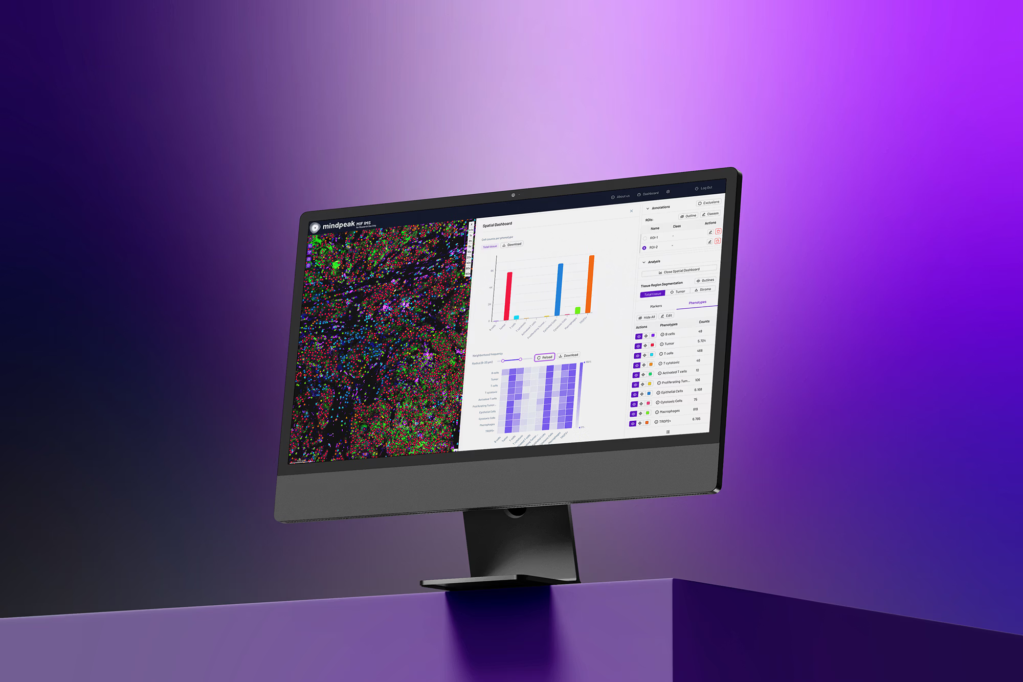
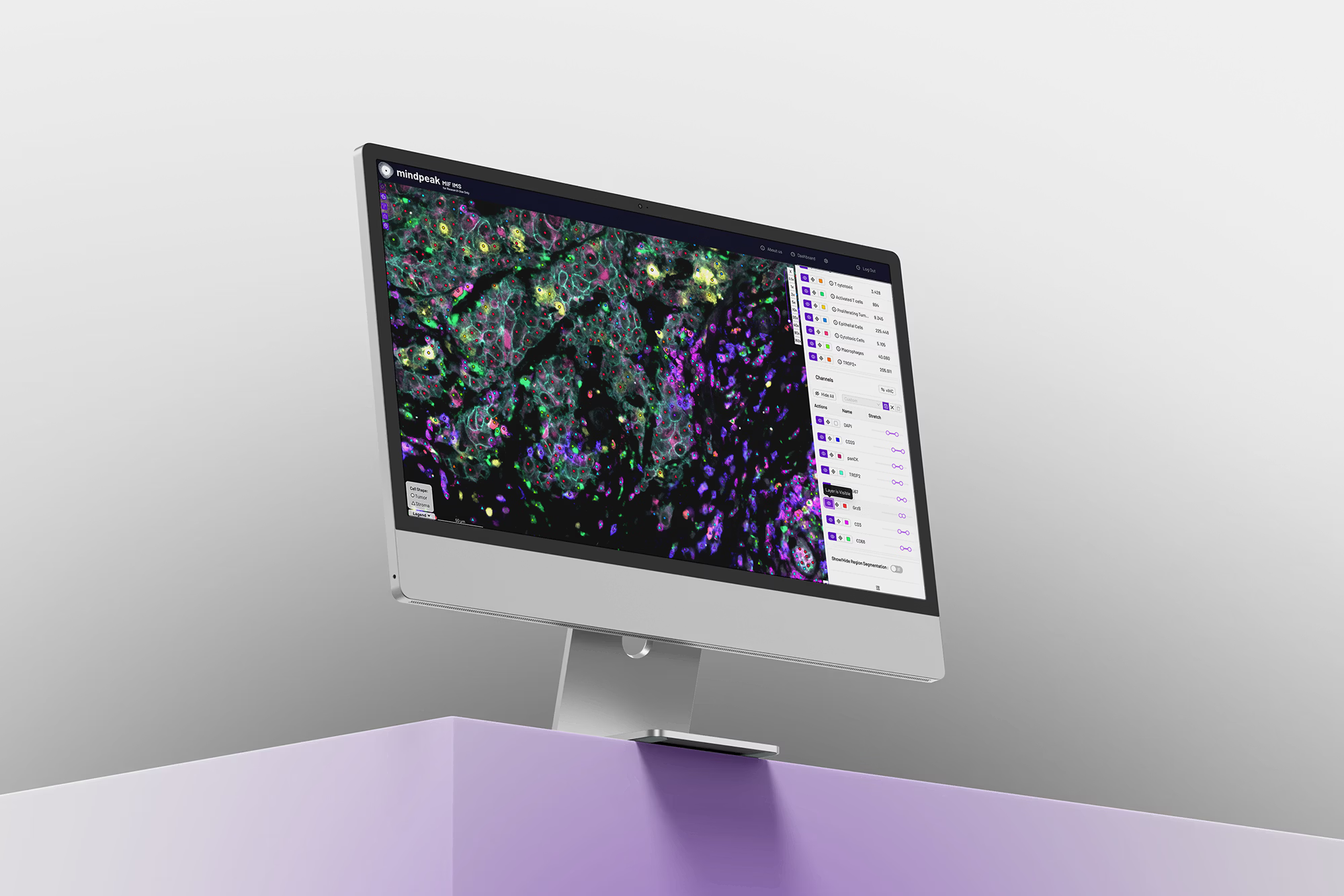
Reveal the tissue biology invisible to the human eye with AI-powered spatial analysis.
Quantifying spatial organization, cellular phenotypes and tissue morphology to support reproducible research and confident decision-making.
Spatial biology was the discovery. Making it routine is what comes next.
Everything Spatial Biology Needs to Drive Confident Decisions
Turn-Key — No Bioinformatician Handoff
A cloud-based mIF image-analysis platform that pairs an image management system with a multiplex Foundation Model. Go from image to client-ready phenotyping with no scripting and no pipeline customization, removing the wait on image-analysis scientists and bioinformatics resources.

Clinical-Grade QC & Audit Trail
A pathologist-centric, gated workflow that mirrors a diagnostic sign-out:
(1) check image quality on visual features,
(2) AI runs only once the image is accepted,
(3) subjective pass/fail review of the AI results.
Every step is traceable and reproducible across operators and samples.

Foundation-Model AI, Publication-Ready Output
The PhenoScout foundation model is pre-trained on 30+ immune and cancer biomarkers and every cell morphology: elongated, amorphous, cobblestone, round, where standard models fail. Single-image spatial analysis (neighborhood-frequency matrix, 0–50 µm) and publication-ready readouts (counts, density, %) meet the RUO publishability bar.
Transforming Spatial Biology Across the Research Ecosystem

For Translational Researchers & Spatial Biologists
Standardized cell classification and spatial insights — without building or maintaining an analysis pipeline.
Pre-trained models that generalize across antibody panels and chemistries, so results are comparable study to study.
From image to single-cell phenotypes and tissue neighborhoods, with explainable, reviewable outputs.
Runs on the multiplex and brightfield slides you already acquire — no custom development.

For BioPharma & Drug Development Teams
Sharper patient stratification — turn every patient image into objective, quantitative spatial signatures that strengthen enrollment decisions, de-risk programs, and support leaner, faster trials.
More from every slide — automated cell-level phenotyping and tissue-context analysis extract the full quantitative and spatial signal already on each scan, so limited tissue goes further and a single dataset can answer multiple questions in parallel.
Decision-ready spatial readouts — reproducible, quantitative outputs that support biomarker validation and companion-diagnostic development across ADC and immuno-oncology programs.
For CROs & Core Labs
High-throughput, walk-away analysis that scales — proven on whole-slide images and large TMAs.
"Zero-friction" cloud access from anywhere, with a pay-what-you-analyze credit model (1 credit = 1 biomarker channel).
Predictable delivery timelines through consistent, automated, reproducible workflows across scanners and operators.

For Academic Research
Bring foundation-model spatial analysis to your lab without a dedicated bioinformatics team.
Flexible access tiers and storage to match project scope, from pilot to multi-centre programme.
From Slide to Spatial Insight in Five Steps with the ZEISS - Mindpeak End to End Workflow


Ready to Accelerate Your mIF Program?
Multiplex IF + AI is transforming biomarker strategy. With PhenoScout you can detect patients others miss, drive smarter therapy decisions, predict efficacy through spatial insight, and discover next-generation combination paths - at routine scale.
Join the researchers, CROs and pharma teams already working with Mindpeak and ZEISS to bring spatial biology from research to routine.
FAQ
Everything you need to know about Phenoscout
PhenoScout accepts multiplex immunofluorescence (mIF) and brightfield images, including cyclic stainings of 20+ channels, and supports images from all major spatial platforms (e.g. RareCyte Orion, Akoya PhenoImager HT, Lunaphore COMET). AI analysis currently runs on the PhenoScout mIF model, with a generic brightfield (IHC) model available as an add-on; support for further modalities is already planned.
No. PhenoScout is a turn-key, off-the-shelf solution. Pre-trained models run on upload, so there's no pipeline to build, no algorithms to tune, and no bioinformatics queue to wait in.
Two pre-trained mIF models — mIF-membrane (for membrane/cytoplasm markers such as HER2, CD8, panCK, CD68) and mIF-nucleus (for nuclear markers such as Ki67, FOXP3) — plus ready-to-use brightfield IHC-membrane and IHC-nucleus models. All are built on the peakFrontier foundation model and the mIF model is trained on 30+ immune and cancer biomarkers.
PhenoScout is cloud-native and browser-based, accessible anywhere, anytime. An on-premise (dockerized, in-network) deployment is available for teams that require image and result data to remain within their own infrastructure upon request.
PhenoScout integrates with leading imaging hardware (including the ZEISS Axioscan 7 / SlideStream workflow) and with laboratory IT systems (LIMS / IMS), from launching results in a separate viewer to embedding them directly in third-party IMS viewers.
PhenoScout AI is For Research Use Only — not for use in diagnostic procedures. It is built to GxP-ready standards, with a full audit trail for every image ensuring traceable, reproducible analysis. Mindpeak is ISO 13485 (QMS) certified, GDPR-compliant and made in Germany, and also develops a separate line of CE-IVD clinical products.
Need more clarity?
Our team is ready to walk through implementation details.